General information
BETTER DIAGNOSIS ENABLES EXCELLENT SURGICAL RESULTS
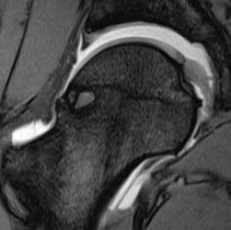
When preparing for hip joint surgery, it is essential to know the condition of the cartilage in the area of the femoral head and the acetabulum, as well as the condition of the labrum, in order to be able to determine a favourable arthroscopic access route, for example, with reference to the local extent of any joint damage that has been identified.
In order to obtain a clear picture of the local extent and size of any joint damage using magnetic resonance imaging, it is advantageous to fill the hip joint with a contrast agent and subject it to tensile stress during the examination.
Advantages
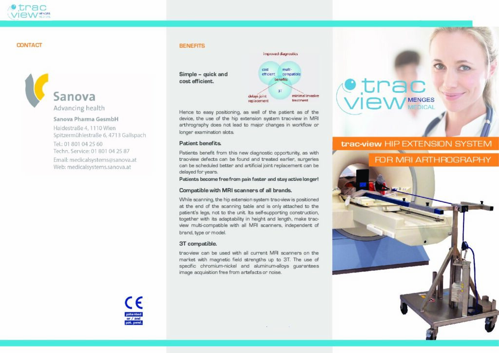
THE HIP EXTENSION SYSTEM FOR PERFECT
MR ARTHROGRAPHY OF THE HIP JOINT SIMPLE, FAST AND COST-EFFECTIVE
The simple application of trac-view enables a quick and smooth examination process. The additional time required compared to conventional MR arthrography is only 2 to 3 minutes.
COMPATIBLE WITH ALL MR DEVICES
The trac-view hip extension system is placed at the foot end of the patient table and is not connected to the MR system. This means it can be used with all MR systems from a wide range of manufacturers.
3T-COMPATIBLE
trac-view can be used with both 1.5 Tesla and 3 Tesla MR systems. A special chrome-nickel steel alloy enables interference-free image acquisition.
Technologie
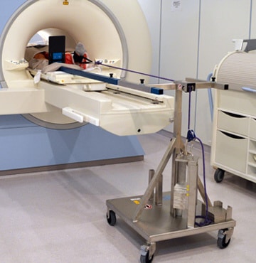
Structure and function
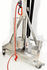
The trac-view hip extension system consists of an integrated central pulley for a cable pull and a support plate that can be locked in two positions (for the right and left leg) in the horizontal plane.
The hip joint of the limb being examined is loaded with a weight of 15 or 18 kilograms using a standard orthopaedic splint and the cable pull system.
To prevent pelvic tilt and the resulting loss of traction, the healthy leg is positioned on the angled support plate. The use of the pulley and support plate ensures that the applied traction is transferred to the hip joint being examined without loss, thus achieving the best possible distension.
Investigation procedure
MR hip arthrography with trac-view
EASY, QUICK, COST EFFECTIVE.
Trac-view® is an easy-to-use diagnostic aid that enables accurate MR imaging of the hip joint in a simple and cost-effective manner.
Following arthrography, the patient is positioned on the MR table in a matter of minutes and an ankle splint is applied to the foot of the limb to be examined. In the next step, Trac-view® is placed at the end of the MR table and the ankle splint is connected to a cable pull system with a weight load of
15 to 20 kilograms.
Before traction is applied, the foot on the opposite side must be positioned on a support plate to prevent pelvic tilt and the associated loss of strength. Without affecting the patient, the joint space is expanded by the applied tensile force, enabling an assessment of the central parts of the hip joint. With the help of this method, lesions on the joint cartilage and pathological changes on the acetabular labrum and the ligamentum capitis femoris can be detected accurately for the first time.
After the examination, Trac-view® can be dismantled in just a few steps and is immediately ready for the next examination. Depending on the sequence protocol, examination times of between 20 and 30 minutes are possible.
Young women and men with hip problems benefit most from the use of Trac-view®. Thanks to improved radiological diagnostics, these patients can be offered a minimally invasive treatment option as an alternative to prosthetic surgery, which restores the joint and delays the need for joint replacement by years.