General information
IMAGE REPRESENTATION IN THE 21ST CENTURY
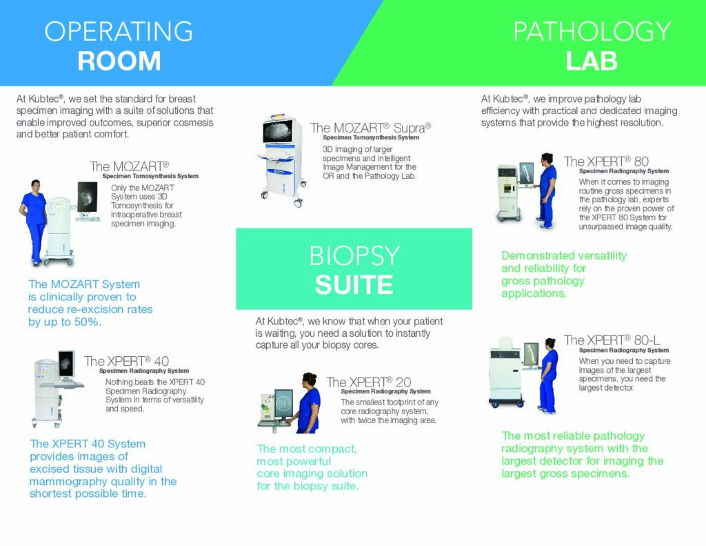
MOZART is the world’s first specimen radiography system that offers both 2D and 3D image viewing, enabling comprehensive analysis of excised breast tissue.
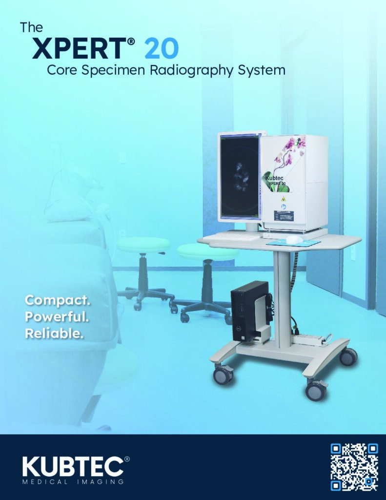
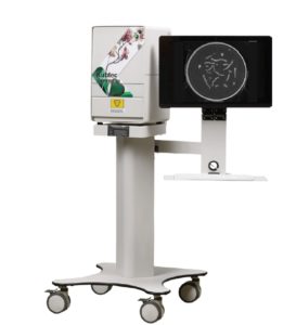
The XPERT 20 is ideal for limited space. The system is available as a stand-alone system or optionally on a mobile cart.
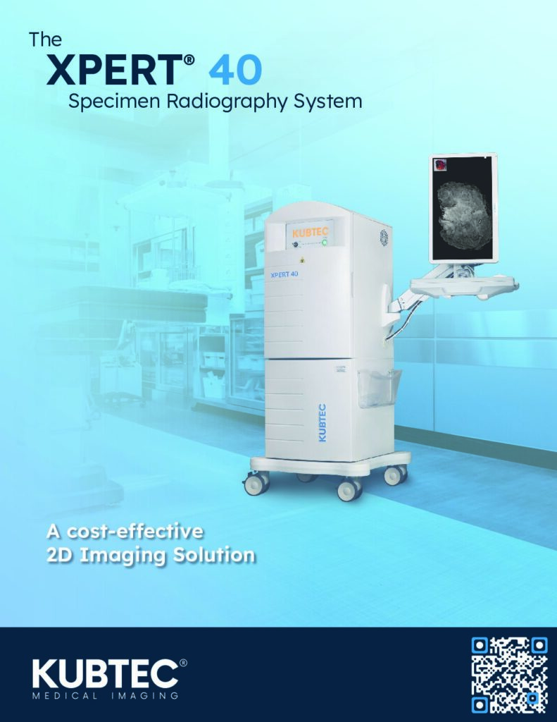
The XPERT 40 is the most versatile device for use in specimen radiography. Its compact, mobile design makes it easy to transport between the biopsy suite and the operating theatre.
Advantages
Advantages of Mozart with Tomospec
- Reduction in repeat surgery rates
- Reduction in patient recalls
- Ensuring improved resection margin assessment with comprehensive 3D sample imaging
- Increasing patient confidence and satisfaction
- Eliminating the need to generate multiple images of a sample from different angles
- Reducing the duration of a procedure
- Increasing the volume of procedures per day
Features of all systems
- Compact and mobile for use in radiology, pathology and surgery
- Fast image display in a few seconds
- Wireless connection
- DICOM-compliant image acquisition and analysis software
- Send multiple images to the PACS with just one click
- No separate sample container required
- Optionally available: easy-to-use touchscreen monitor
MOZART® with TomoSpec®
THE WORLD’S FIRST SAMPLE IMAGING SYSTEM WITH TOMOSYNTHESIS
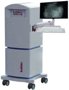
MOZART® with TomoSpec® is the world’s first sample radiography system that offers both 2D and 3D image viewing, enabling comprehensive analysis of excised breast tissue. Thanks to the possibilities offered by tomosynthesis, MOZART® with TomoSpec® is able to provide an unprecedented 3D view of the specimen – including views that are not possible with conventional 2D imaging devices. With its superior 2D/3D image display capabilities and extremely fast image rendering, MOZART with TomoSpec is the most powerful X-ray imaging system for assessing resection margins.
TomoSpec®
PATENT-PENDING TOMOSYNTHESIS TECHNOLOGY FOR EVALUATING RESECTION MARGINS
TomoSpec, Kubtec’s groundbreaking technology, is the first of its kind in digital specimen tomosynthesis. TomoSpec provides you with excellent 3D images of a resected breast tissue specimen, the ability to examine specimens of varying thicknesses, and includes K-VIEW® image generation for a comprehensive 2D view of multiple acquired layers in higher resolution.
With TomoSpec, you no longer need to continuously position samples at different angles to confirm margins. The high-resolution 3D data provides more detail and greater accuracy without sacrificing time compared to 2D imaging.
MOZART® with TomoSpec requires no warm-up phase and features automatic calibration tools to ensure fast acquisition of detailed, high-resolution 2D and 3D images – every time.
FEATURES
- Compact and mobile for use in radiology, pathology and surgery
- Slim, ergonomic design with height-adjustable monitor
- Anti-microbial surfaces
- No separate sample container required
- Easy-to-use touchscreen monitor available
- DICOM-compliant image capture and analysis software
- Send multiple images to PACS with just one click
- Super-fast image display in a matter of seconds
XPERT 20/40
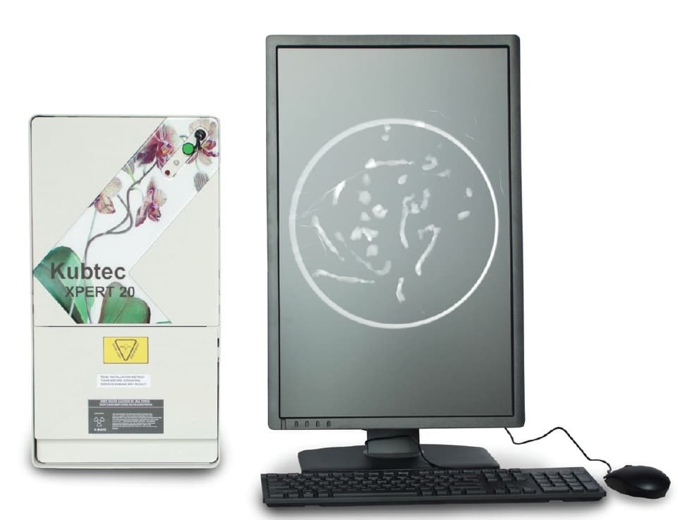
XPERT™ 20
Ideal for limited space and to improve the efficiency of your biopsy suite: the XPERT 20 enables you to provide high-quality treatment for your patients, even in the procedure room.
The system is available as a stand-alone system or optionally on a mobile cart, helping you to increase patient throughput while reducing anxiety and restlessness. The system provides you with high-resolution images in seconds, allowing you to identify even the smallest microcalcifications.
XPERT™ 40
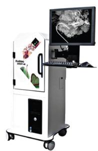
The XPERT 40 is the most versatile device for use in sample radiography. Its compact, mobile design makes it easy to transport between the biopsy suite and the operating theatre. A 50kV, 1.0 mA X-ray source even allows dense tissue to be penetrated and shows fine details within seconds.
An integrated optical camera is also available as an option for comparing 2D images side by side. The XPERT 40 is ideal for biopsies and surgical samples.
DIGICOM® DICOM-compliant software
HIGH-QUALITY IMAGES FOR HIGH-QUALITY TREATMENT
Kubtec’s dynamic and user-friendly DIGICOM software offers you comprehensive image analysis tools for every type of radiography application.
In addition to tools such as system control, annotations, measurements, zoom and image filters, DIGICOM software includes many other tools for analysing your images and specific areas. DIGICOM allows you to save high-resolution images in multiple formats and send multiple images with or without annotations to PACS with just one click.
The XPERT series digital X-ray devices require no warm-up time and feature automatic calibration tools to ensure fast, high-resolution image capture at all times.